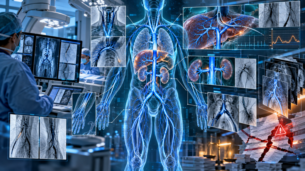
Few areas of interventional radiology require more coding precision than head and neck vascular interventions. These procedures are among the most technically challenging to code due to their complex vascular anatomy, intricate selective catheterization pathways, and the frequent combination of diagnostic and therapeutic services performed during a single encounter. Coders must accurately identify vascular territories, distinguish diagnostic angiography from interventional procedures, correctly report embolization, angioplasty, or stent placement, and apply evolving CPT® coding and bundling guidelines. Case examples are an excellent way to evaluate and strengthen critical coding knowledge while closing gaps in understanding. The following case follows the treatment of a symptomatic patient with severe right internal carotid artery stenosis who underwent comprehensive cerebral angiography before successful carotid artery stenting with distal embolic protection. Because these procedures often combine diagnostic evaluation, selective catheterization, embolic protection, angioplasty, and stent placement, coders must carefully apply CPT® guidelines to determine which services are separately reportable and which are bundled into the definitive intervention.
VASCULAR INTERVENTIONS CASE: CAROTID ARTERIOGRAM AND INTERVENTION
Indications: The patient is an 81-year-old gentleman with a history of transient ischemic attack and recently current episodes of left facial droop, and is now referred for angiographic evaluation and percutaneous intervention with distal embolic protection.
Procedure:
1. Thoracic aortic arch arteriogram.
2. Selective bilateral intracerebral and extracranial carotid arteriogram.
3. Percutaneous right internal carotid artery intervention with distal embolic protection.
4. Radiologic supervision and interpretation.
Conclusions:
- Type I aortic arch without significant atherosclerotic disease or lesions at the origins of the great vessels.
- 80% stenosis in the proximal portion of the right internal carotid artery with an ulcerated plaque.
- Successful percutaneous intervention, with placement of a 10 x 40 mm SMART stent across the right carotid artery bifurcation reducing the stenosis to 20% residual preserving TIMI grade III flow.
Procedure Note: Informed consent was obtained. The patient had been treated with clopidogrel for more than 7 days prior to admission. He was prepped and draped in the usual manner. The right groin was infiltrated with 1% lidocaine. A #5 French sheath was placed in the right femoral artery. Thoracic aortic arch angiogram was performed through a 5 pigtail catheter. Selective bilateral carotid arteriogram was performed with a JB1 diagnostic catheter in each common carotid artery. For percutaneous intervention, adjunctive bivalirudin therapy was administered. The 5 French sheath was exchanged for a 6 French 19 cm Destination sheath over a TAD wire. Hemostasis in the arterial access site was achieved with a ProGlide device after angiographic confirmation of appropriate femoral artery anatomy. 200 cc of Visipaque contrast was used for the procedure.
Findings:
- Thoracic aortic arch angiogram. The patient has a type I aortic arch. The origins of the innominate, left common carotid, and the left subclavian arteries were visualized. No significant occlusive lesions were appreciated. No significant atherosclerotic disease was noted in the ascending aorta and aortic arch.
- Selective intracerebral and extracranial carotid arteriogram. a. The left common carotid artery is free of significant stenosis. It gives rise to the left external carotid and internal carotid arteries. There is a 20% stenosis near the origin of the left internal carotid artery. The left external carotid artery is free of significant disease. b. There is no significant occlusive disease noted in the mid and distal portions of the left internal carotid artery. c. The left anterior cerebral and middle cerebral arterial circulation appears to be intact. d. The right common carotid artery originates from the innominate artery. It is free of significant disease. It gives rise to the right external carotid and internal carotid arteries. e. The proximal portion of the right internal carotid artery has a long concentric up to 80% stenosis. There is also an area of ulceration. There is no angiography evidence of thrombus. The TIMI flow grade into the distal right internal carotid artery is III. f. Right external carotid artery is free of significant disease. g. The right anterior cerebral and middle cerebral artery circulation appears intact.
- Percutaneous carotid artery intervention. A TAD wire was advanced through the JB1 diagnostic catheter into the right external carotid artery without difficulty. The JB1 diagnostic catheter as well as the 5 French arterial sheath were exchanged for a 6 French 90 cm Destination sheath, which was advanced over the wire into the right common carotid artery. Subsequent angiogram confirms the position of the arterial sheath in the mid portion of the right common carotid artery. The proximal right internal carotid artery stenosis was once again studied for multiple projections. A 7 mm ANGIOGUARD filter device was advanced across the lesion into the straight portion just before the petrous section of the right internal carotid artery. The filter was deployed without difficulty. Next, a 4 x 40 mm Viva balloon was positioned into the index lesion and inflated to 8 atmospheres for 20 seconds. The patient had transient bradycardia with balloon inflation. Next, a 10 x 40 mm SMART stent was then positioned over the index lesion. It was deployed without difficulty. The stent crosses the right common carotid artery bifurcation and covers the lesion well. Post-stent dilatation was then performed with an Aviator 5 x 20 mm balloon, which was positioned into the stented segment and inflated to 12 atmospheres for 15 seconds. Final angiography revealed approximately 20% residual stenosis in the stented segment of the proximal right internal carotid artery. There was no obvious plaque shifting into the right external carotid artery which has intact flow. Final angiography revealed no evidence of dissection. The final TIMI flow grade was III.
Complications: The patient tolerated the procedure well. There were no in-lab complications. Estimated blood loss: Less than 20 cc.
Assessment/Recommendation: Has undergone successful revascularization of a severe symptomatic stenosis in the proximal portion of the right internal carotid artery with good angiographic result. He will be monitored overnight. If stable he will be ready for discharge on the next hospital day. He will complete a 30 day course of antiplatelet therapy with clopidogrel. In addition, he will be treated with 81 mg daily of aspirin. Cardiovascular risk factors will continue to be aggressively pursued.
CODE ASSIGNMENTS AND RATIONALE
37215 Transcatheter placement of intravascular stent(s), cervical carotid artery, open or percutaneous, including angioplasty, when performed, and radiological supervision and interpretation; with distal embolic protection
36223-59 Selective catheter placement, common carotid or innominate artery, unilateral, any approach, with angiography of the ipsilateral intracranial carotid circulation and all associated radiological supervision and interpretation, includes angiography of the extracranial carotid and cervicocerebral arch, when performed
Report code 36223 with modifier 59 for catheterization of the left common carotid artery and diagnostic imaging of the left cervical and cerebral carotid circulation. Code 37215 is assigned for the placement of the stent in the right internal carotid artery with the use of a filter for embolic protection. Code 37215 includes ipsilateral carotid catheterization and diagnostic imaging, so it would be inappropriate to code 36223 again.
⚠️Your IR Coding Remains Under Threat, Creating Significant Risk to Your Bottom Line. These Are NOT All the Tips and Tricks Necessary for Success.⚠️
With every dollar of reimbursement counting more than ever in the face of payment decline and complex changes, it’s imperative to make sure your CPT® coding is correct and compliant. Master more coding topics and break down the complexity with our 2026 IR Masterclass: Head and Neck Interventional Radiology Coding webcast on July 15, 2026 at 11:00 am CT (120 minutes). This webcast is an essential training tool for both audio and visual learners.