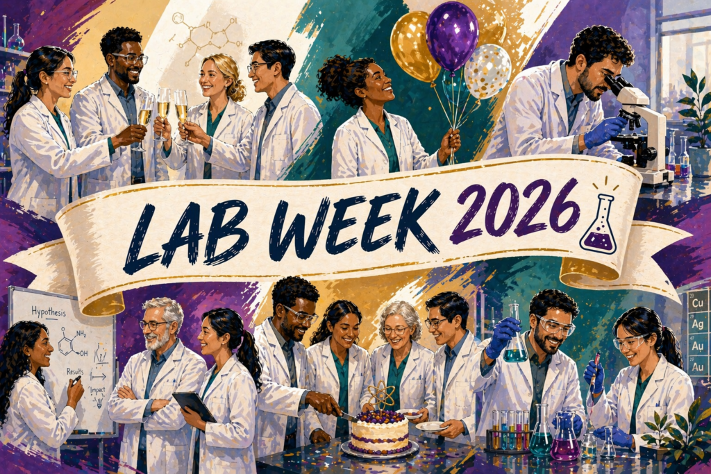
Venous Coding Case Example for Enhanced Understanding

Case studies are great learning tools for coders, providing an array of detail and rationale to enhance understanding. Here, we explore a case study related to venous access for correct coding. By examining case studies like these, coders can ensure success throughout the year.

VENOUS ACCESS CASE: TEMPORARY DIALYSIS CATHETER PLACEMENT

Clinical History: Adult patient with end-stage renal disease, difficult venous access, and nonfunctioning right groin AV graft needs intermediate-term central venous access for hemodialysis. The vascular surgeon is planning for a new AV graft in the left thigh. The referring nephrology service has asked for a tunneled dialysis catheter placement in the right groin.

Procedure Performed: Right femoral tunneled hemodialysis catheter placement. The risks, benefits and alternatives to the procedure were explained to the patient, and informed written consent was obtained. The patient was brought to the interventional radiology suite where a time-out procedure was performed. The patient was placed in the supine position. The right groin was prepped and draped in the usual sterile fashion. Real-time ultrasound was used, and a permanent image stored. Using ultrasound guidance the right common femoral vein was punctured with a micropuncture needle, after infiltration of the skin and deep tissues with local anesthetic. A 50 cm tip to cuff length, 14.5-Frenchdual lumen Vaxcel hemodialysis catheter was inserted. The catheter was placed through a subcutaneous tunnel requiring a second incision. The incision at the femoral vein access site was closed with 4-0 Vicryl suture. The catheter was secured at the skin exit site with 2-0 Prolene suture. There is free aspiration of blood from all ports of the catheter, which were locked with heparin (concentration 1000 units/cc). A sterile dressing was then applied. Postprocedure chest fluoroscopy showed the tip of the catheter at the right atrium, and an image was stored in the medical record.

The pre-existing left femoral catheter was removed in its entirety. A sterile dressing was applied. The patient tolerated the procedure well with no immediate complications. This procedure was performed using ultrasound and fluoroscopy. Impressions:

  1. Ultrasound of the right groin demonstrates patent common femoral vein and compressible.
  2. Successful right femoral tunneled hemodialysis catheter placement with its tip positioned at the right atrium as discussed above.

There is free aspiration of blood from all ports of the catheter. The catheter is ready for immediate use.

CODE ASSIGNMENTS AND RATIONALE
36558Insertion of tunneled centrally inserted central venous catheter, without subcutaneous port or pump; age 5 years or older
+76937Ultrasound guidance for vascular access requiring ultrasound evaluation of potential access sites, documentation of selected vessel patency, concurrent real-time ultrasound visualization of vascular needle entry, with permanent recording and reporting (List separately in addition to code for primary procedure)
+77001Fluoroscopic guidance for central venous access device placement, replacement (catheter only or complete), or removal (includes fluoroscopic guidance for vascular access and catheter manipulation, any necessary contrast injections through access site or catheter with related venography radiologic supervision and interpretation, and radiographic documentation of final catheter position) (List separately in addition to code for primary procedure)

Ultrasound was used to locate a suitable vein and to guide the needle access into that vein. An image of the vein was obtained for a permanent record. A catheter was placed from a central vein (femoral vein) into the right atrium of the heart using fluoroscopic guidance including a permanent image of the final position. The catheter was tunneled from a skin entry site away from the vein.

Although the report documents that a previously existing catheter was removed, there is no documentation that it was a tunneled catheter or that dissection was required to remove it. Therefore, no removal code would be assigned.

Explore more regulatory, policy, and coding knowledge to master compliance and coding with our monthly Radiology Compliance Manager.

Print Friendly, PDF & Email
Facebook
Twitter
LinkedIn

You May Also Like

Leave a Reply

Please log in to your account to comment on this article.

Subscribe

Subscribe to receive our News, Insights, and Compliance Question of the Week articles delivered right to your inbox.

Resources You May Like

Trending News

Happy World Health Day! Our exclusive webcast, ‘2024 SDoH Update: Navigating Coding and Screening Assessment,’  is just $99 for a limited time! Use code WorldHealth24 at checkout.

SPRING INTO SAVINGS! Get 21% OFF during our exclusive two-day sale starting 3/21/2024. Use SPRING24 at checkout to claim this offer. Click here to learn more →